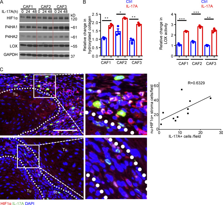
Figure S3.
IL-17 induced HIF1α expression and collagen deposition in human CAFs. (A) Western blot analysis of IL-17A–treated CAFs for 24 and 48 h. (B) Hydroxyproline assay and LOX assay for IL-17A–treated and untreated (Ctrl) human CAFs (48 h). n = 3 technical repeats. Error bars represent ± SEM; *, P < 0.05; **, P < 0.01; ***, P < 0.001 by t test. CAFs for A and B were from three independent human cutaneous SCCs, and data are representative of three independent experiments. (C) Immunofluorescence analysis of IL-17A (green)–producing cells and HIF1α (red) expression in two representative cutaneous SCCs. Scale bar, 100 μm. St, stroma; Tu, tumor islets. Dotted lines are used to show the main boundary of tumor islets and stroma areas. Graph shows correlation of nuclear HIF1α-positive cells (nu-HIF1α+) and IL-17A–producing cells in random stroma areas (40× magnification) of 10 human cutaneous SCCs.

