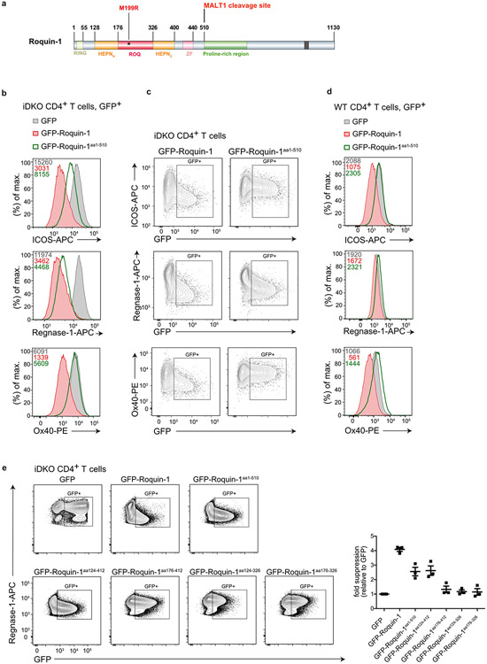
Extended Data Fig. 5 ∣. Dissecting protein domains of Roquin-1 sufficient for cooperative target regulation with Regnase-1.
(a) Schematic representation of Roquin-1 domain organization with indication of M199R mutation. iDKO (b, c) or WT (d) CD4+ T cells were retrovirally transduced with GFP, GFP-Roquin-1 or GFP-Roquin-1aa1-510 constructs. Histograms of flow cytometry analysis of ICOS, Regnase-1 or Ox40 expression, as indicated, in GFP+ cells with indication of respective geometric MFI value. (c) Contour plots of histograms depicted in (b). (e) iDKO CD4+ T cells were retrovirally transduced with the constructs encoding GFP, GFP-Roquin-1 or GFP-Roquin-1 mutant proteins. Flow cytometry analysis of Regnase-1 expression and quantification of fold suppression of Regnase-1 expression level in GFP+ cells relative to cells expressing GFP control construct. (b–e) Data are representative of n = 3 independent experiments. (e) Data are presented as mean +/− SEM of n = 3 independent experiments.

