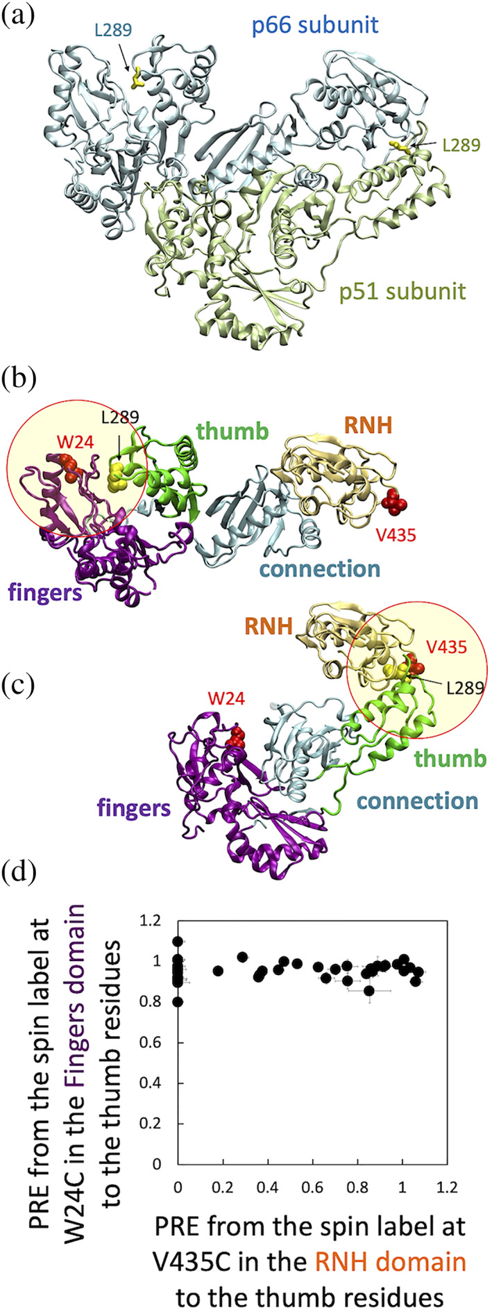
FIGURE 1.

(a) Location of residue L289 in the p66/p51 crystal structure, (b, c) two hypothesized thumb domain orientations based on the p66/p51 crystal structure, (d) PRE, defined as intensity ratio of I para /I dia (Section 5), to thumb residues from the MTSL‐labeled W24C fingers domain relative to those from MTSL‐labeled V435C RNH domain. In (a), p66 subunit (light cyan) and p51 subunit (light green), with L289 side chain in each subunit (yellow sticks), are shown with ribbon presentation using PDB 1DLO. 26 Panels (b) and (c) show RT domains in the same orientation as that of panel (a), in a way to have one p51 and one RNH to generate one p66 monomer. In (b), the p66 subunit in p66/p51 structure is shown with highlight of fingers domain residue 24 (red van der Waal spheres). In (c), the p51 subunit in p66/p51 structure and the RNH domain in p66 are shown with highlight of RNH domain residue 435 (red van der Waal spheres). In (b) and (c), fingers‐palm, thumb, connection, and RNH domains are colored with purple, green, cyan, and light orange, respectively. The circles indicate a ~20 Å radius, at which proton PRE is sensitive (Note, since these circles are drawn in two‐dimensions, the distance is only approximate). In (d), normalized PREs from residue 24 to thumb residues are plotted on the Y‐axis while PREs from residue 435 to the same thumb residues are plotted on the X‐axis
