Fig. 7.
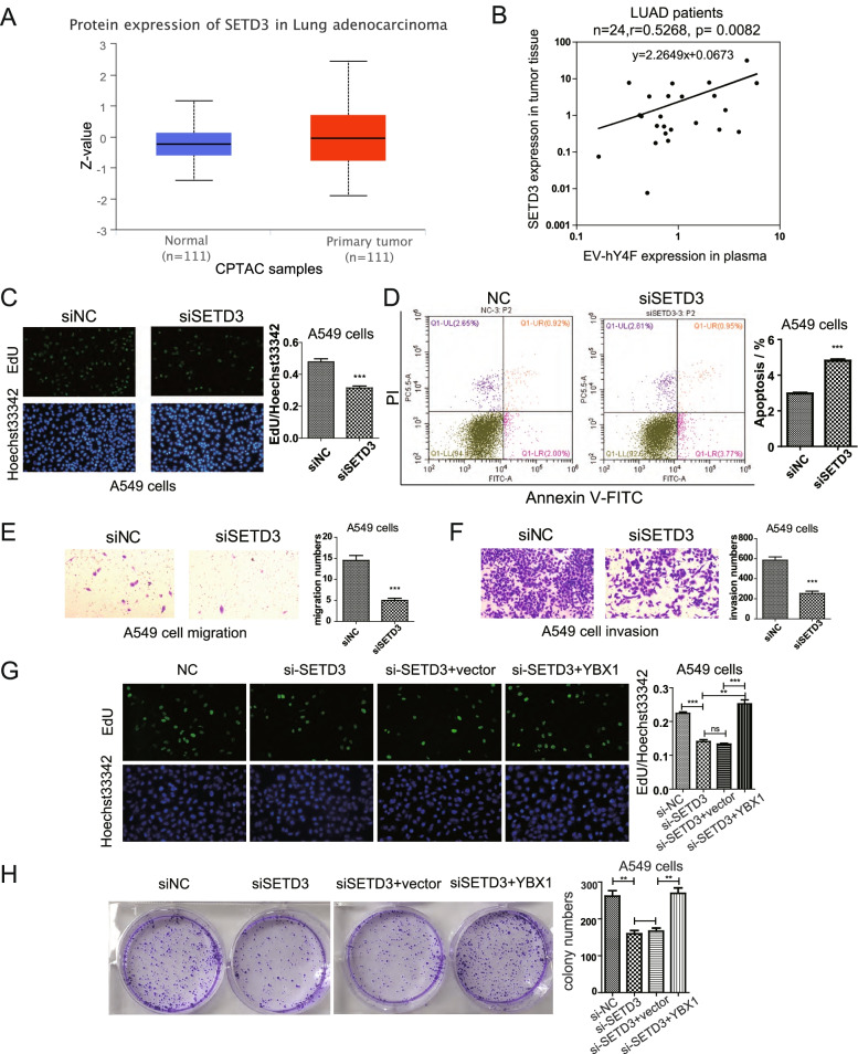
SETD3 promotes lung cancer cell proliferation and migration in A549 cells through YBX1/EVs-hY4F axis. A Levels of SETD3 protein in tissue from LUAD cancer patients or control subjects according to the CPTAC database (P = 0.0387). B Pearson correlation analysis of plasma EV-hY4F and tumor tissue SETD3 expression levels in lung adenocarcinoma (LUAD) patients (n = 24) detected by qPCR. Transcript levels of SETD3 were normalized by internal control 18S rRNA, and plasma hY4F levels were normalized by external control cel-miR-39-3p. C EdU assay was performed to assess the effect of SETD3 knockdown on A549 cell proliferation (at 24 h after transfection). D Flow cytometry using Annexin V-FITC/PI staining was performed to analyze apoptosis of A549 cells transfected with SETD3 siRNA (at 24 h after transfection). The effect of SETD3 knockdown on A549 (E) migration and (F) invasion was examined by transwell assay (at 24 h after transfection). G EdU assay was performed to assess the effect of YBX1 co-transfection with siRNA targeting SETD3 on A549 cells (at 48 h after transfection). H Colony formation assay with YBX1 and siSETD3 co-transfected A549 cells (at 7 d after transfection). Data from three independent experiments are shown as the mean ± SD (error bars). *P < 0.05, **P < 0.01, ***P < 0.001 (Student’s t-test). NC: negative control

