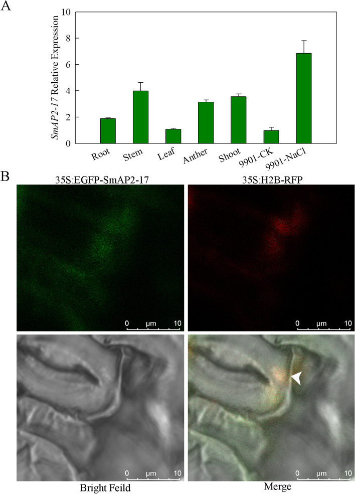
Fig. 3.
Expression pattern and subcellular localization of SmAP2-17 protein. a Expression patterns of SmAP2-17 in the roots, stems, leaves, anthers, and shoots of S. matsudana and under salt stress were measured using qRT-PCR. b Subcellular localization of the SmAP2-17 protein. The 35S:EGFP-SmAP2-17 fusion construct and the nucleus localization marker 35S:H2B-RFP construct were co-transformed into tobacco epidermal leaves. The arrowhead indicates the merged signal (yellow) with EGFP (green) and RFP (red) co-located in the nucleus. Scale bar, 10 μm

