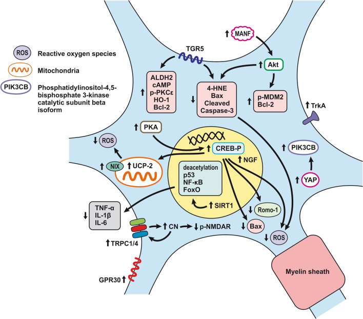
Fig. 7.
Neuronal protective mechanisms after SAH. SAH-induced neuroprotection involves decreased cell apoptosis, necrosis, neuronal regeneration, oxidative injury, inflammation, and removal of damaged mitochondria. Attenuation of neuronal apoptosis is supported by molecules up-regulated by TGR5, MANF, Akt, SIRT1, UCP-2, and GPR30. Increased expression of TGR5 leads to up-regulation of anti-apoptotic protein levels including cAMP, p-PKCε, ALDH2, HO-1, Bcl-2, and down-regulation of pro-apoptotic proteins such as 4-HNE, Bax, and cleaved caspase-3. SAH induced. MANF leads to activation of Akt, resulting in up-regulation of p-MDM2 and Bcl-2 and down-regulation of pro-apoptotic molecules. Up-regulation of SIRT1 contributes to the deacetylation of acetylated-forkhead box O, acetylated-NF-κB, and acetylated-p53, thus attenuating the oxidative, inflammatory, and apoptotic pathways. Increased UCP2 levels lead to decreased ROS generation. Moreover, an increase in PKA expression phosphorylates CREB, which results in UCP2 upregulation and downregulates Bax and Romo-1. Up-regulation of TrkA, NGF, and YAP/PIK3CB contribute to neuronal growth and regeneration and BBB maintenance (increased expression of occludin and claudin-5 induced by YAP/ PIK3CB pathway). Dephosphorylation of NMDAR via up-regulation of TRPC1/4 and subsequent CN protects neurons from excitotoxicity

