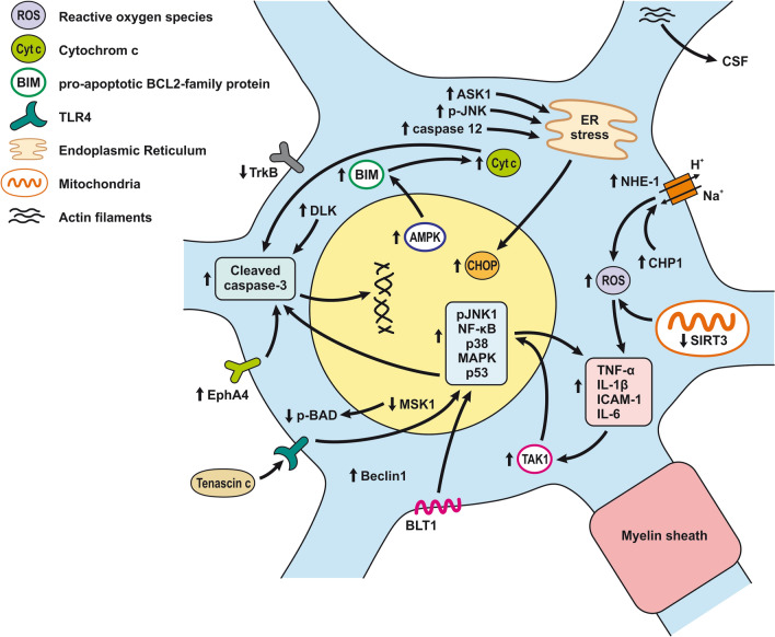
Fig. 8.
Neuronal injury and inflammation after SAH. Schematic illustration of pathophysiological cascades leading to the development of inflammation and neuronal injury after SAH. Induction of NF-κB via activation of TLR4 (tenascin C), TAK1 (TNF-α and IL-1β), and BLT1 (LTB4) leads to overexpression of proinflammatory molecules such as TNF-α and IL-1β as well as ICAM-1. In addition to pro-inflammatory molecules, NF-κB, JNK, and p38 activate the mitochondrial apoptotic pathway and upregulate cleaved-caspase3 resulting in DNA fragmentation and neuronal death. Prolonged activation of AMPK leads to induction of Bim, which promotes Cyt c release from mitochondria and subsequent maturation of caspase-3. Increased expression of Eph receptor A4 (EphA4) and DLK also contributes to caspase-dependent neuronal death. Down-regulation MSK1 leads to decreased phosphorylation of Bcl2-associated agonist of cell death (Bad) and thus promotes neuronal death. Decreased activation and phosphorylation of TrkB reduces neuronal protection and thus increases neuronal apoptosis. Similarly, increased expression of CHP1 activates Na+H+-exchanger 1 (NHE1), which contributes to oxidative stress resulting in neuronal death. Down-regulation of SIRT3 contributes to the increased generation of ROS. The endoplasmic reticulum stress-related apoptotic proteins such as C/EBP homologous protein (CHOP), caspase-12, ASK1, and p-JNK are increased following SAH. The induction of these proteins contributes to neuronal apoptosis during the SAH

