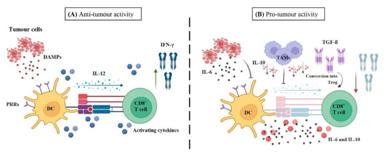
Figure 3.
Anti- and pro-tumour microenvironment of DCs and CD8+ T cells. (A) Antitumour microenvironment: Antigen presentation by DCs via MHC-I to CD8+ T cells is depicted. For T cell activation to take place, apart from antigen presentation, co-stimulatory signals (DAMPs released by tumour cells, which are recognised by the PRRs of DCs and thus activated) and activating cytokines (represented by the blue round shapes) are necessary. Accordingly, activated DC can also promote CD8+ T cell by releasing IL-12 which induces IFN-γ production by CD8+ T cells. (B) Immunosuppressive microenvironment. However, factors such as tumour cells-derived IL-6 production or TAMs-derived IL-10 production (involved in inhibiting DC-derived IL-12) can negatively influence maturation, migration, differentiation and hence, function of DCs, downregulating them and give rise to a pro-tumour microenvironment. TGF-β, another factor present in TME, is able to convert effector T cells into Treg, creating an immunosuppressive environment. DAMPs: Damage-associated molecular patterns; PRRs: pattern recognition receptors; DC: dendritic cell; IL-12: Interleukin-12; IFN-γ (interferon gamma); TAMs: Tumour-associated macrophages.

