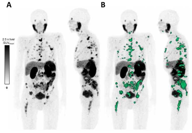
Figure 1.
(A) MIP (maximum intensity projection) of [68Ga]Ga-PSMA-11 PET in an advanced mCRPC patient. (B) Semi-automatic tumor segmentation by Syngo.via (Enterprise VB 60, Siemens, Erlangen, Germany). SUV windowing was set from 0 to 2.5 times SUVmean of the liver. The delineated tumor volume is shown in green.

