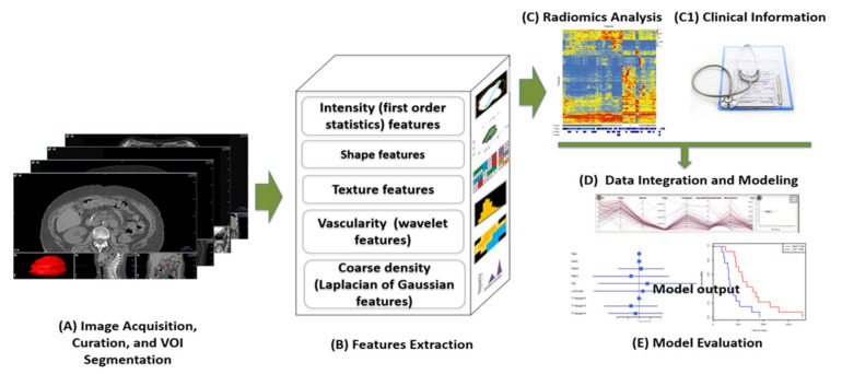
Figure 3.
In a typical radiomics workflow, medical images are acquired and curated and volumes of interest (VOIs) such as pancreatic tumors are segmented (A). From the segmented VOI images, hundreds to thousands of radiomic features are then be extracted (B). After conducting preliminary radiomics analysis such as feature selection (C) and possibly adding clinical and biological information (C1), all features can be integrated through advanced statistical and/or machine learning methods to develop predictive models (D). The model accuracy and robustness can then be evaluated on validation and testing datasets (E).

