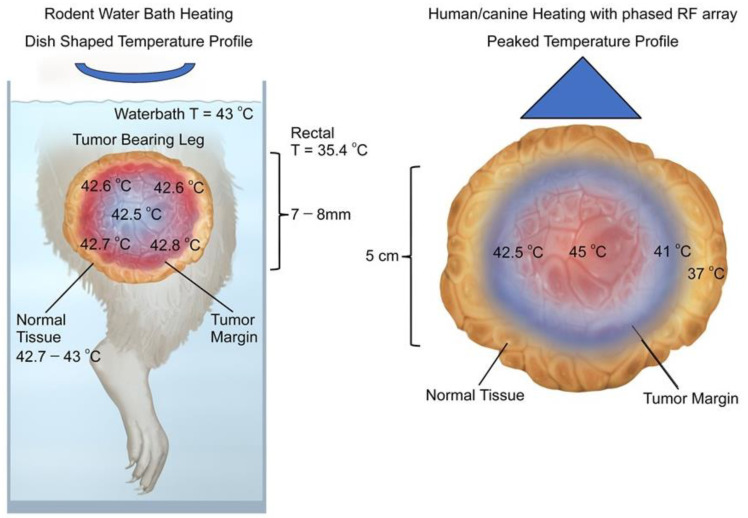
Figure 1.
Schematic comparison of temperature distributions for rodent water bath heating vs. temperatures seen in canine and human tumors heated with radiofrequency or microwave devices. (Left Panel) Temperature distributions in rodent tumors heated with water baths tend to be relatively uniform [profiles are dish-shaped], with highest temperatures at the margin of the tumor, while intra-tumoral temperatures are slightly cooler and relatively uniform. Depicted data are taken from a paper by O’Hara et al., where detailed intra-tumoral temperatures were documented using micro-thermocouples [72]. Although not shown in color for clarity, the whole leg is at elevated temperature. This is described numerically at the left side of the figure. (Right Panel) Temperature distributions in human and canine sarcomas heated with phased radiofrequency devices have a peaked temperature distribution in which the temperatures closer to the center are higher than those at the tumor edge. Typically, some surrounding normal tissue is heated to mild temperatures, as depicted. Note also that maximum intra-tumoral temperatures are higher than what is seen in rodent tumors. This is a schematic representation of non-invasive thermometry obtained in human sarcomas [79,80,81].

