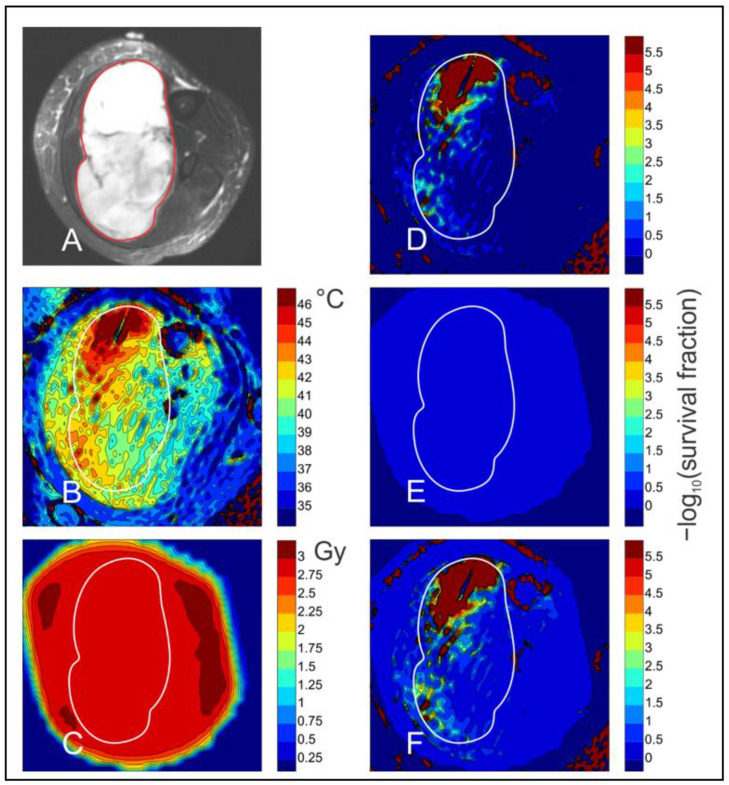
Figure 4.
Imaging and simulation of combined HT and radiation treatment of a sarcoma. Images show a cross-section through a human patient’s calf. Simulations are based on results of Loshek et al. [139] for dependence of survival fraction of Chinese hamster ovary cells on doses of combined radiation and heating, together with results of Sapareto and Dewey [61] for the dependence of thermal dose on temperature. The period of heating was 54 min. Details of the simulation are provided in Text S1. (A) Diffusion weighted MRI image of thigh cross-section. Tumor region is outlined in red and transferred to other images. (B) Temperature distribution in tissue during hyperthermia, obtained by non-invasive MRI thermometry. (C)Radiation dose derived from treatment plan. (D) Predicted cell kill from HT alone. All cell kill values are expressed in terms of −log10 (survival fraction). (E) Predicted cell kill from radiation alone. (F) Predicted cell kill from combined HT and radiation.

