Figure 2.
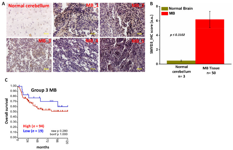
High SMYD3 expression is associated with reduced survival in MB. (A) Immunohistochemical (IHC) staining of normal human cerebellar tissue and MB tissues (magnification 40×; scale bar, 50 µm) (B) Q score analysis was performed using (staining intensity_Intden/no. of positive cells) ImageJ software to quantify SMYD3 protein expression. Staining for IHC analysis of MB (n = 50) and controls (n = 3) was performed per field (4×/per specimen) (C) Kaplan–Meier survival plot showing the overall survival probability of Group 3 MB patients with high (red) or low (blue) SMYD3 transcripts.

