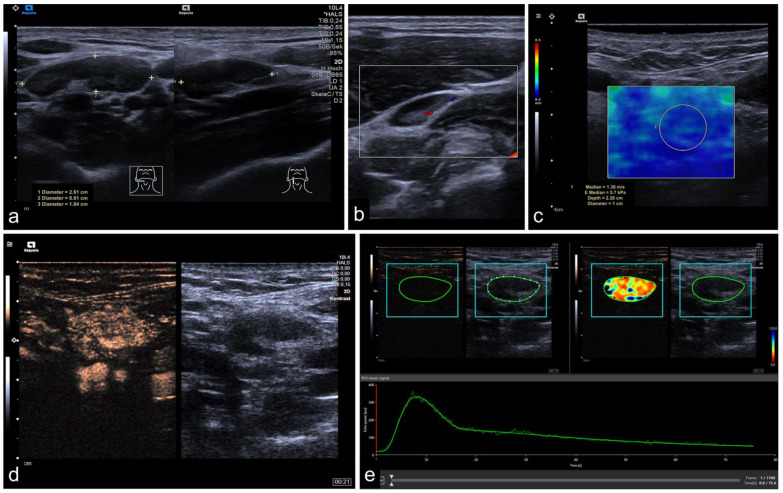
Figure 1.
Representative image of mpUS workflow. (a) Long-axis and short-axis diameter measured in dual mode, (b) color-coded Doppler US depicting macrovascularity, (c) SWE measured by circular ROI revealed low tissue stiffness (1.38 m/s), (d) CEUS image 21 s after contrast injection with regular enhancement, and (e) RAW data perfusion analysis with demarcation margins (blue) and lesion ROI (green) showing the corresponding time-intensity curve and color-coded map. The CLN was confirmed as benign.

