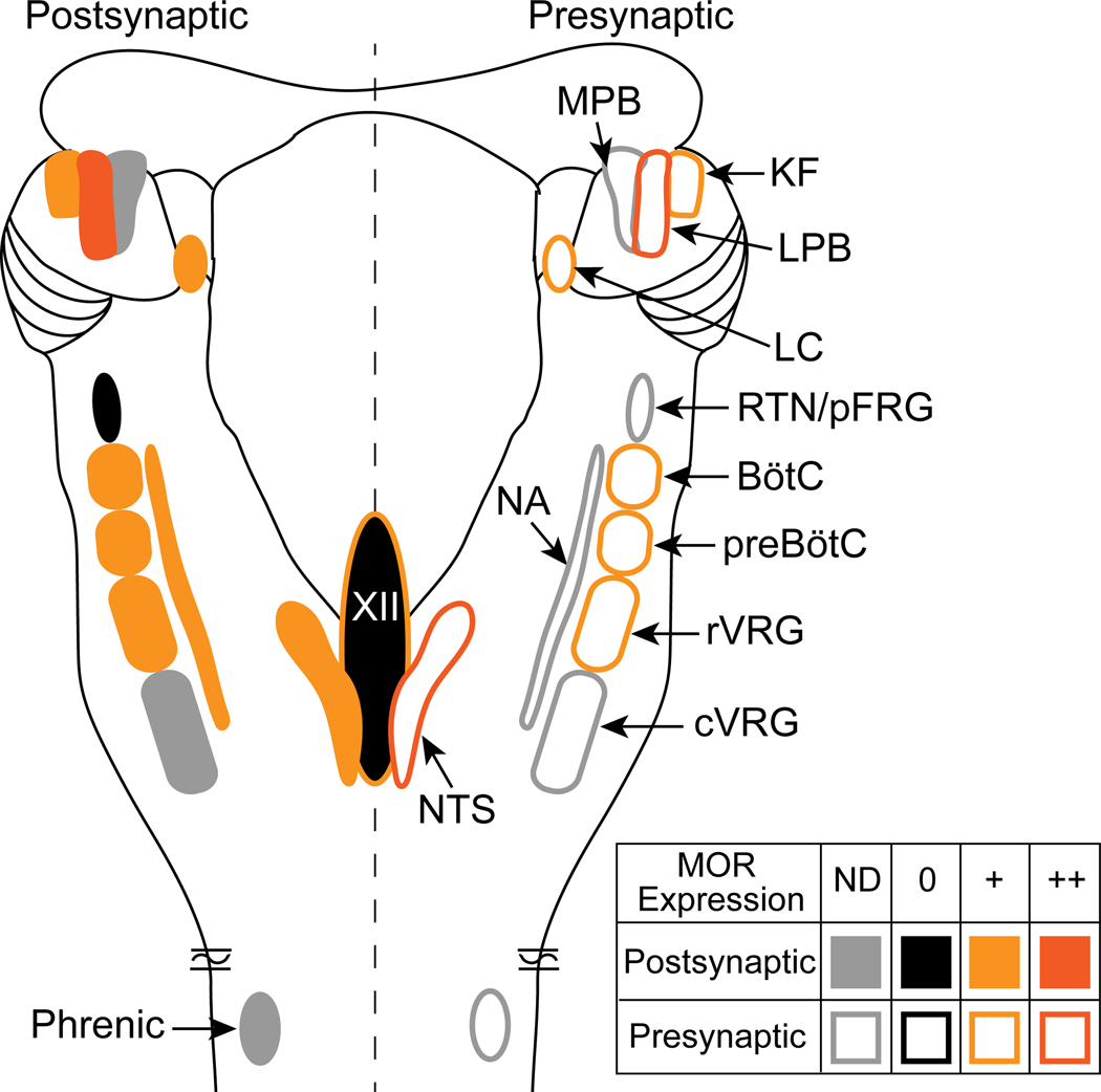
Figure 2.
Mu opioid receptor distribution in the pontomedullary respiratory network. Dorsal view of a rodent brainstem with bilateral pontomedullary respiratory structures is depicted. Mu opioid receptor expression is indicated as postsynaptic (somatodendritic, left side of brainstem, filled symbols) or presynaptic (axon terminals, right side of brainstem, outlined symbols). In each area, mu opioid receptors are expressed (+, orange), highly expressed (++, dark orange), not expressed (-, black) or not determined (ND, gray). Details are in the text (section 3. Brainstem mechanisms of opioid-induced respiratory depression). Abbreviations and references: MOR, mu opioid receptor; KF, Kölliker-Fuse (Levitt et al., 2015); LPB, lateral parabrachial area (Chamberlin et al., 1999); LC, locus coeruleus (Bradaia et al., 2005; Levitt and Williams, 2012); RTN/pFRG, retrotrapezoid nucleus/parafacial respiratory group (Mulkey et al., 2004); BötC, Bötzinger complex (Lonergan et al., 2003); preBötC, preBötzinger complex (Lonergan et al., 2003; Gray et al., 1999; Sun et al., 2019; Bachmutsky et al., 2020b); rVRG, rostral ventral respiratory group (Lonergan et al., 2003); cVRG, caudal ventral respiratory group; NTS, nucleus of the solitary tract (Aicher et al., 2000; Poole et al., 2007); XII, hypoglossal motor nucleus (Lorier et al., 2010); NA, nucleus ambiguous (Erbs et al., 2015); Phrenic, phrenic motor neurons.

