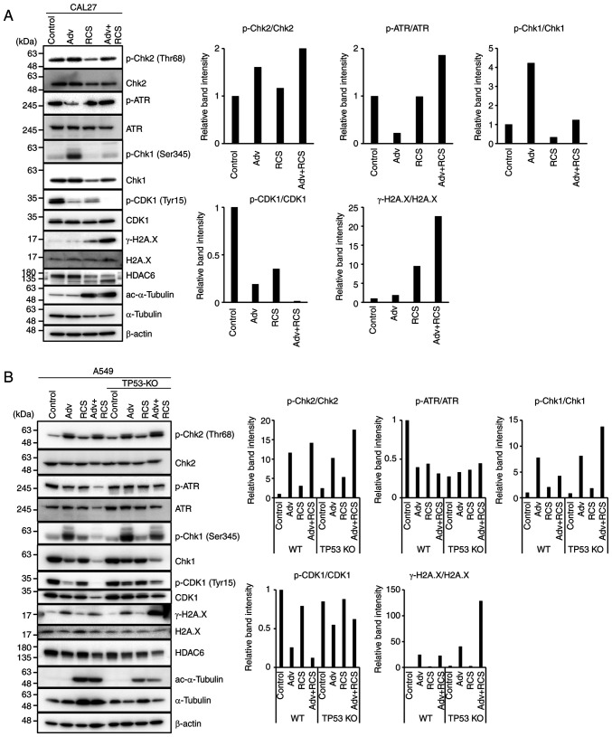
Figure 4.
Ricolinostat suppresses phosphorylation of Chk1 and further suppresses p-CDK1 when co-administered with adavosertib. (A) CAL27 cells and (B) TP53-WT and TP53-KO A549 cells were treated with Adv (0.5 µM), RCS (5 µM), and Adv+RCS for 24 h (CAL27 cells) or 48 h (A549 cells), and then the expression of DNA damage response-related proteins (p-Chk2, Chk2, p-ATR, ATR, p-Chk1, Chk1, p-CDK1, CDK1, and γ-H2A.X) was assessed by western blotting. To assess the inhibitory effect of HDAC6 by RCS, the level of acetylated (ac)-α-tubulin was monitored. Expression of β-actin was assessed as the loading control. The relative band intensity of each phosphorylated protein was calculated and summarized at the right. Representative data of three independent experiments are shown. Adv, adavosertib; RCS, ricolinostat; WT, wild-type; Chk, checkpoint kinase; ATR, ATR serine/threonine kinase; CDK1, cyclin-dependent kinase 1.

