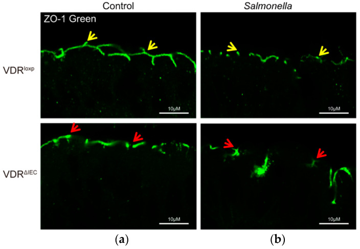
Figure 3.
ZO-1 distribution before and after Salmonella infection in mouse colon. (a) ZO-1 distribution in the colon of VDRloxp colon and VDRΔIEC mice. The yellow arrows indicate the “zipper” structure of ZO-1 in the apical side of the colon, and red arrows show the disrupted ZO-1 due to the intestinal epithelial VDR deletion in VDRΔIEC mice. (b) Disrupted ZO-1 distribution after Salmonella infection in the colon of VDRloxp and VDRΔIEC mice. VDRloxp and VDRΔIEC mice aged 6–8 weeks were infected with Salmonella and sacrificed 8 h post-infection, as described in our previous study [27,28].

