Figure 2.
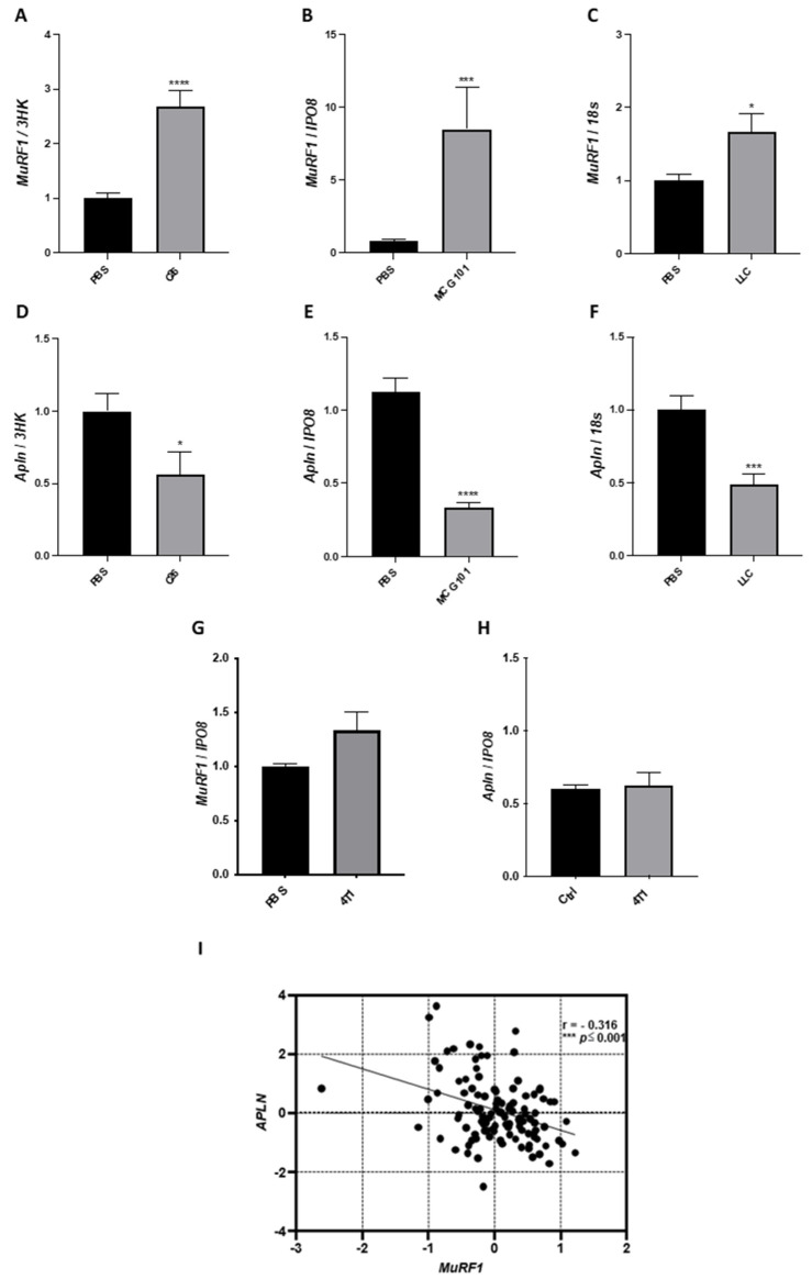
In the atrophied muscle of all the three animal models of cancer cachexia and cancer patients, apelin expression falls when MuRF1 increases. Q-PCR shows that mRNA levels of the ubiquitin ligase MuRF1 are highly induced in the TA of mice carrying the tumor C26 (A) or MCG101 (B) or LLC (C). Unpaired t-test, **** p ≤ 0.0001 (A); Mann–Whitney test, *** p ≤ 0.001 (B). Unpaired t-test, * p ≤ 0.05 (C), n = 7–10 (A–C). Apelin (Apln) expression is drastically decreased in TA from mice bearing C26 (D) or MCG101 (E) or LLC (F). Unpaired t-test, * p ≤ 0.05 (D), **** p ≤ 0.0001 (E), *** p ≤ 0.001 (F), n = 7–10. The mRNA levels of MuRF1 are not induced in the TA of mice carrying the non-cachectic tumor 4T1 (G) and apelin expression is not altered in this tumor model (H). 3HK, three housekeeping genes (TBP, IPO8, and Gusb), IPO8 or Importin 8 and 18S or 18S ribosomal RNA, were used to normalize the data. Unpaired t-test, not significant, n = 5. In rectus abdominis muscles from cancer patients, as soon as MuRF1 increases, apelin decreases (I). The scatter plot shows the inverse correlation between the expression of apelin and MuRF1. These expression data are log2-transformed. Spearman’s test, with correlation index (r) = −0.316, *** p ≤ 0.001, n = 115.

