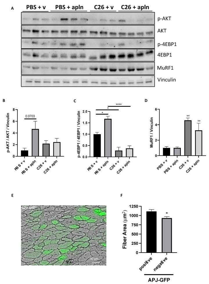
Figure 6.
Muscles from mice with cancer cachexia display apelin resistance. Proteins from TA muscles of C26-carrying mice injected with preproapelin-expressing AAV9 were analyzed in Western blot for p-AKT, AKT, p-4EBP1, 4EBP1, MuRF1, and vinculin as loading control (A). Quantitations for p-AKT/AKT/vinculin (B), p-4EBP1/4EBP1/vinculin (C), and MuRF1/vinculin (D) are shown. Ordinary one-way ANOVA, Tukey’s multiple comparison test, * p ≤ 0.05, ** p ≤ 0.01, **** p ≤ 0.0001, n = 3–4. The APJ-GFP-encoding plasmid was electroporated for 14 days in mice that were injected with C26 tumor cells the day after. A representative image is shown (E). Scale bar, 50 μm. At sacrifice, TA were dissected, frozen, and cut and the mean fiber CSA of APJ-GFP-expressing fibers was found bigger than that of adjacent non-expressing ones (F). Unpaired t-test, Mann–Whitney test, * p ≤ 0.05, seven mice and 89 fibers.

