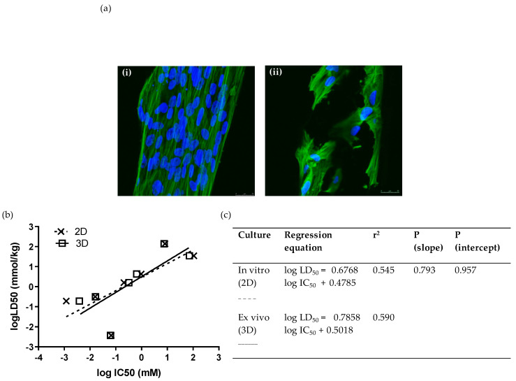
Figure 2.
Ex vivo cytotoxicity screening of 7 selected chemicals on 3D scaffolds by means of the WJSC-MTS assay: (a) Growth and morphology of WJSCs in the 3D microenvironment of the inert polystyrene (PS) scaffold. Panels show representative composite confocal micrographs of WJSCs growing on the periphery of the microtubules comprising the scaffold meshwork (i) in the presence of growth medium (GM) only (control), or (ii) in GM containing a concentration of NaF close to its IC50. Each composite image was derived by merging 10 confocal micrographs taken across the tube diameter at 1.5 um steps (z-series). Mag = 40×, Mag bar = 25 uM; blue = DAPI nuclear staining; green = phalloidin staining of the actin cytoskeleton. (b,c) Comparison of linear regressions of human fetal WJSCs cultured on conventional tissue-culture-treated plastic (polystyrene (PS)) surface (2D culture), and in three-dimensional inert scaffolds with a rectangular mesh structure (PS inserts).

