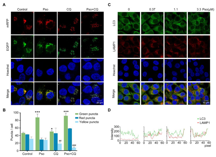
Figure 5.
Psoralen promotes the autophagic flux by facilitating the fusion of the autophagosome and lysosome. (A) L02 cells were induced with sodium oleate (100 μM) for 24 h, transfected with the mRFP-EGFP-LC3 plasmid, and then treated with psoralen (3.3 μM) or CQ (10 μM) for 24 h. Nuclei were stained with Hoechst 33258 (scale bar = 20 μm). (B) Quantification of puncta per cell. (C) L02 cells were induced with sodium oleate (100 μM) for 24 h and treated with psoralen at 0.37, 1.1, 3.3 μM for 24 h, and stained with Hoechst 33258, anti-LC3, or anti-LAMP1 antibodies (scale bar = 10 μm). (D) Colocalization efficiency of LC3 and LAMP1 was quantified by line scan analysis (60 pixels with two ends on the membrane) by observing the overlap of fluorescence intensity peaks across the contours of multiple L02 cells (n ≥ 3 cells). All values were expressed as the mean ± SD from three independent experiments. * p < 0.05, *** p < 0.001 vs. sodium oleate-induced group. ## p < 0.01, ### p < 0.001, vs. psoralen treated group. Abbreviations: CQ, chloroquine; LAMP1, lysosomal associated membrane protein 1; Pso, psoralen.

