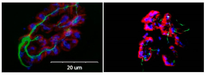
Figure 3.
Micrograph showing close coupling of presynaptic vesicles and terminal branches with postsynaptic receptors. Presynaptic terminal branches are stained green, presynaptic vesicles are stained blue and postsynaptic receptors are stained red. Note the greater complexity of nerve terminal branching in aged NMJ.

