Figure 6.
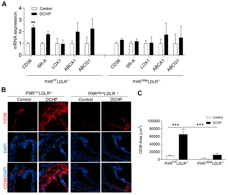
Deficiency of myeloid PXR reduces DCHP-induced CD36 expression in macrophages and atherosclerotic lesions of PXRΔMyeLDLR−/− mice. Four-week-old male PXRF/FLDLR−/− and PXRΔMyeLDLR−/− littermates were fed a low-fat diet and treated by oral gavage with 10 mg/kg body weight of DCHP or vehicle control daily for 12 weeks. (A) Total RNAs were isolated from fresh isolated peritoneal macrophages of PXRF/FLDLR−/− and PXRΔMyeLDLR−/− mice, and the expression levels of indicated genes were analyzed by QPCR (n = 4–6, ** p < 0.01). (B) Representative images of immunofluorescence staining of CD36 (Red) in the aortic root of PXRF/FLDLR−/− and PXRΔMyeLDLR−/− mice (Scale bar = 100 µm). The nuclei were stained with DAPI (Blue). (C) Quantification analysis of CD36 staining area in the aortic root of PXRF/FLDLR−/− and PXRΔMyeLDLR−/− mice (n = 5, *** p < 0.001). Data are represented as mean ± SEM.

