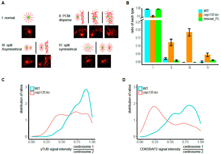
Figure 7.
Imbalanced MTOC functions in cep135 KO cells in interphase. (A) The typical patterns of MT regrowth in WT and cep135 KO cells (red: MT, green: γ-tubulin). MT regrowth was allowed after cold treatment for 15 s. (B) Quantification of differently patterned microtubule structures in regrowth assays as exemplified in (A), I–IV. The graph shows mean values ± SD from three independent experiments. (C,D). The two distinct γ-tubulin and CDK5RAP2 signals in interphase cells with separated centrosomes were quantified, and their ratios (higher/lower value) were determined. The plot shows the distribution of ratios ranging from 1 (equal signals) to 0 (extreme asymmetry between the two signals). A total of 180 WT and 300 KO cells were measured.

