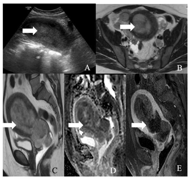
Figure 11.
A 52-year-old female with a large cell neuroendocrine tumor of the endometrium. (A) Transverse ultrasound image of the uterus demonstrates thickened endometrium (arrow), (B) axial T2 weighted image, (C) sagittal T2 weighted image, (D) sagittal apparent diffusion coefficient map, and (E) sagittal fat-saturated post-contrast T1 weighted MRI images demonstrate a hypoenhancing tumor in the endometrium (arrow) with restricted diffusion.

