Figure 1.
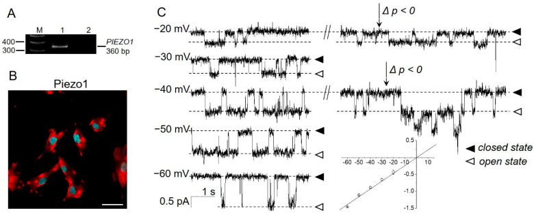
Presence of Piezo1 in eMSCs. (A) RT-PCR analysis revealed the presence of PIEZO1 mRNA. M—size marker, Line 1—primers specific for PIEZO1 amplified the PCR product of the expected size (360 bp), Line 2—RT-PCR negative control in which reverse transcriptase was omitted. Shown is cropped gel with enchanced contrast. (B) Immunofluorescent staining detected Piezo1 proteins (red channel) in eMSCs. No staining of the cells was observed after incubation of the cells with only fluorescent secondary antibodies (Supplementary Figure S2). Cell nuclei were counterstained with DAPI (blue channel). The scale bar is 50 µm. (C) The single-channel activity of Piezo1 induced by selective chemical Piezo1 agonist Yoda1 (10 µM in the pipette solution). Representative current recordings at different membrane potentials are shown. Holding membrane potentials are indicated near current traces, closed and open states indicate the baseline (“zero” current) and Piezo1 open state, respectively. Note that the application of “negative” pressure (p < 0, suction, indicated by the arrows) further increased the activity of the Yoda1-induced channels. The mean I–V relationship corresponded to single-channel conductance of 23.2 ± 1.3 pS (n = 6).

