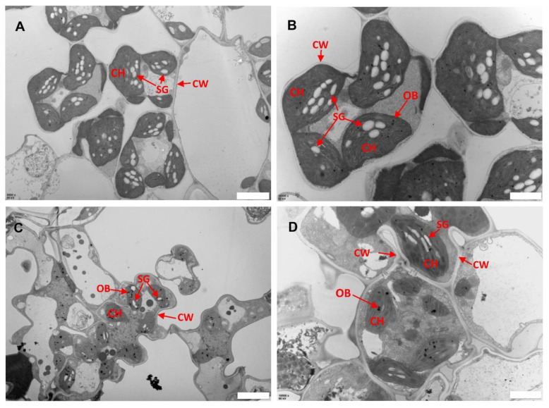
Figure 4.
Transmission electron micrographs of leaf sub-cellular structure of WT and llm9428. (A,B) Subcellular structure of leaf chloroplast of WT, and llm9428 (C,D) at flowering stage under a transmission electron microscope. Where CW: cell wall, CH: chloroplast; SG: starch granules and OB: osmiophilic body. Scale bar: 2 μm in (A,C) and 5 μm in (B,D).

