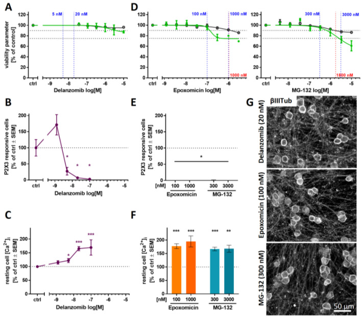
Figure 7.
Attenuation of P2X3 signaling and microtubule reorganization as potential PI class effects. The PIs delanzomib (A–C,G), epoxomicin, and MG-132 (D–G), representing different PI classes, were investigated regarding their effects on various test endpoints. (A,D) The compounds’ effects on neurite area and viability were assessed with the standard PeriTox test. Horizontal dashed lines at 90% and 75% indicate the cytotoxicity threshold and the neurite effect threshold, respectively. Vertical dashed lines indicate the lowest cytotoxicity-inducing concentration (red) and concentrations further used for Ca2+ imaging experiments (blue). (B,C,E,F) Sensory neurons (>DoD38) were pre-treated with the test compounds for 24 h before Ca2+ imaging experiments were performed. (B,E) The number of cells responding to stimulation with the P2X3-specific agonist α,β-methylene ATP (1 µM) was assessed. (C,F) Baseline fluorescence, indicating the resting intracellular Ca2+ concentration ([Ca2+]i) was quantified for whole sensory neuron cultures. (A–F) Data are given as % of untreated control cells and are expressed as the mean ± SEM of at least 3 biological replicates. *, p < 0.05; **, p < 0.001; ***, p < 0.0001. (G) After differentiation of >38 days, sensory neurons were exposed to the PIs for 24 h, fixed, and stained for βIII-tubulin. Representative immunofluorescence images are shown. The scale bar is given in the images. Further details and quantification of cells with circular βIII-tubulin staining are given in Figure S12.

