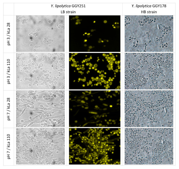
Figure 6.
Microscopic images of Y. lipolytica LB and HB cells cultured under specified conditions (1 column) observed under 1000× magnification in white light (left and right panels) and under fluorescent microscope (central panels). Pictures are representative for the cell morphology starting from 24 h of culturing. Identical fields are shown in left and central panel. Images are representative for a larger number of observations.

