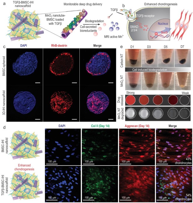
Figure 3.
Deep delivery of soluble factors for enhancing stem-cell chondrogenesis. (a) Schematic diagram of drug releasing and monitoring in the TGFβ–BMSC–IHI nanoscaffold. (b) The effective and homogeneous delivery of TGF-β3 further improved the chondrogenic differentiation of BMSC through Smad pathways. (c) Fluorescence microscopic images demonstrating the deep and homogeneous delivery of model bio-macromolecular drug (Dex–RhB) in the MnO2 NT Dex–RhB-templated IHI nanoscaffold as compared to the control BMSC spheroids incubated with free Dex–RhB. Scale bar: 200 μm. (d) Immunostaining results on chondrogenic markers (Col II, labeled with green, Aggrecan, labeled with red) demonstrated significant enhancement of chondrogenesis of BMSC differentiated in the TGFβ–BMSC–IHI nanoscaffold compared to the BMSC–IHI nanoscaffold. (e) Time-dependent biodegradation of MnO2 NTs in cell culture without the addition of any external trigger. Carbon nanotube (CNT) was used as a negative control and no noticeable degradation was observed. The stoichiometrical release of T1 active Mn2+ enabled the monitoring of MnO2 NTs degradation and drug release, which was confirmed by a direct correlation between the amount of released drug (indicated by red fluorescence) and T1 MRI intensities detected from the nanoscaffold.

