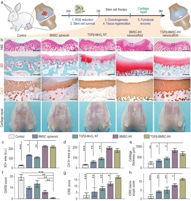
Figure 6.
Accelerated cartilage repair by transplantation of 3D-IHI nanoscaffold. (a) A schematic diagram illustrating the long-term (3-month) cartilage-regeneration process. (b) The in vivo cartilage regeneration was characterized through H&E, Safranin O staining, Col II immunochemistry staining, as well as macroscopic views. Zoom-out scale bars: 2 mm, zoom-in scale bars: 200 μm. (c)–(h) Quantifications of cartilage thickness (by H&E staining) (c), cellular components (by Safranin O staining) (d) and ECM components (by Col II immunostaining) (e). Results of International Cartilage Repair Society (ICRS) macroscopic (f) and histologic scores (g) indicated significantly improved defect repair qualities in the TGFβ–BMSC–IHI nanoscaffold group. The reduced Osteoarthritis Research Society International (OARSI) scores revealed the TGFβ–BMSC–IHI nanoscaffold could prevent the deterioration of osteoarthritis (h). These results collectively suggest that improved cartilage regeneration could be achieved through a MnO2 NT-templated cell assembly and homogeneous delivery of TGF-β3. All data are presented as mean ± SD (n = 5). *P < 0.05, **P < 0.01, ***P < 0.001.

