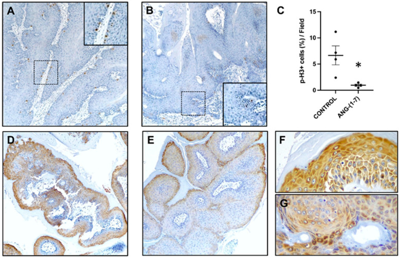
Figure 4.
(A–C): Proliferative status of the oral papilloma from the K14-CreERtam/LSL-K-ras G12D/+ mice treated with vehicle or angiotensin-(1-7). (A) p-H3 expression in a control mouse. The immunostaining is widely extended in the basal layers (inset). (B) p-H3 expression is reduced after angiotensin-(1-7) treatment (detail depicted in the lower inset). (C) p-H3 positive cells were counted and quantified as a percentage of total cells. Angiotensin-(1-7) treated tumors showed a significant reduction of p-H3 labeled cells. Student t-test (n = 4) * p < 0.05. (D–G) pS6 immunostaining in representatives papilloma developed by K14-CreERtam/LSL-K-ras G12D/+ mice treated with vehicle (D,F) or angiotensin-(1-7) (E,G). Level of pS6 in the tumor of a mouse treated with the vehicle was more prominent and showed stronger signal intensity than the angiotensin-(1-7) treatment (E,G). Original magnifications: A,B ×10; D,E ×20; and F,G ×40.

