Figure 3.
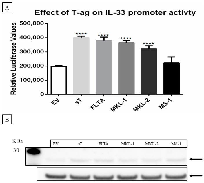
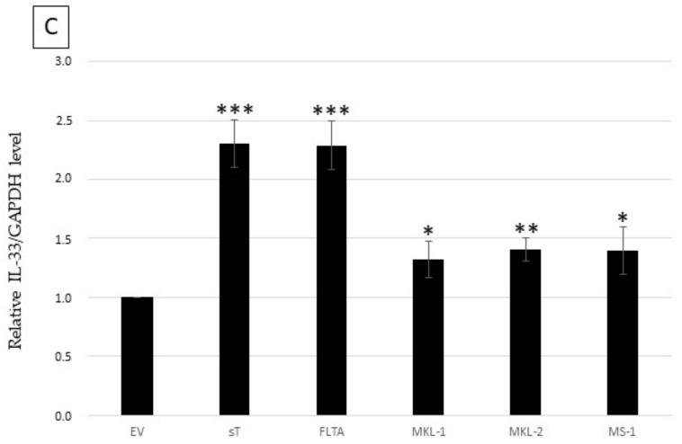
MCPyV T-antigens stimulate IL-33 promoter activity and induce IL-33 expression levels. (A) MCC-13 cells were co-transfected with a luciferase reporter vector driven by the IL-33 promoter fragment spanning the nucleotides −1050/+50, together with an expression plasmid for full-length LT (FLTA), tLT (LTMKL-1, LTMKL-2, LTMS-1), sT, or pcDNA3 (empty vector; EV). Luciferase activity was assessed 24 h after transfection. Each bar represents the average of three independent parallels ± SD. Luciferase values were normalized for total protein in each sample. ** p ≤ 0.01, *** p ≤ 0.001 and **** p ≤ 0.0001. (B) MCC-13 cells were transfected with expression plasmids for LT or tLT, sT, or empty vector, and protein expression was measured at 24 h after transfection. IL-33 expression was normalized with GAPDH. The image is representative for three independent experiments. (C) Ratio of the values obtained by densitometric scanning of the signals obtained with IL-33 and GAPDH antibodies. The ratio for empty vector:GAPDH was arbitrarily set as 1.0 ± standard deviation (* p < 0.05; ** p < 0.01; *** p < 0.001; ****p<0,0001).


