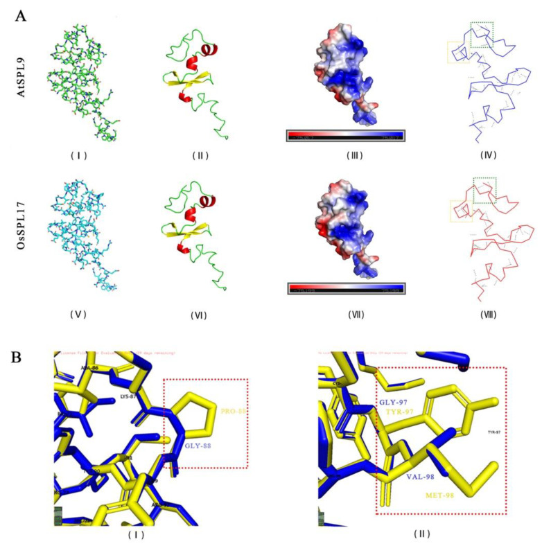
Figure 5.
Comparative analysis of AtSPL9 and OsSPL17 protein structures. (A) The stick structure (A(I,V)), secondary structure (A(II,VI)), surface electrostatic potential (A(III,VII)) and hydrogen bonding structure (A(IV,VIII)) of AtSPL9 and OsSPL17 proteins. (B) Differences in amino acid composition and protein structure between OsSPL17 and AtSPL9. In (A(II,VI)), the red part indicates the α-helix and the yellow part indicates the β-fold. In (A(IV,VIII)), the gray dashed lines indicate hydrogen bonds. In (B), the yellow and blue structures indicate the amino acid arrangement of AtSPL9 and OsSPL17, respectively. PRO: Proline. GLY: Glycine. TYR: Tyrosine. MET: Methionine. VAL: Valine.

