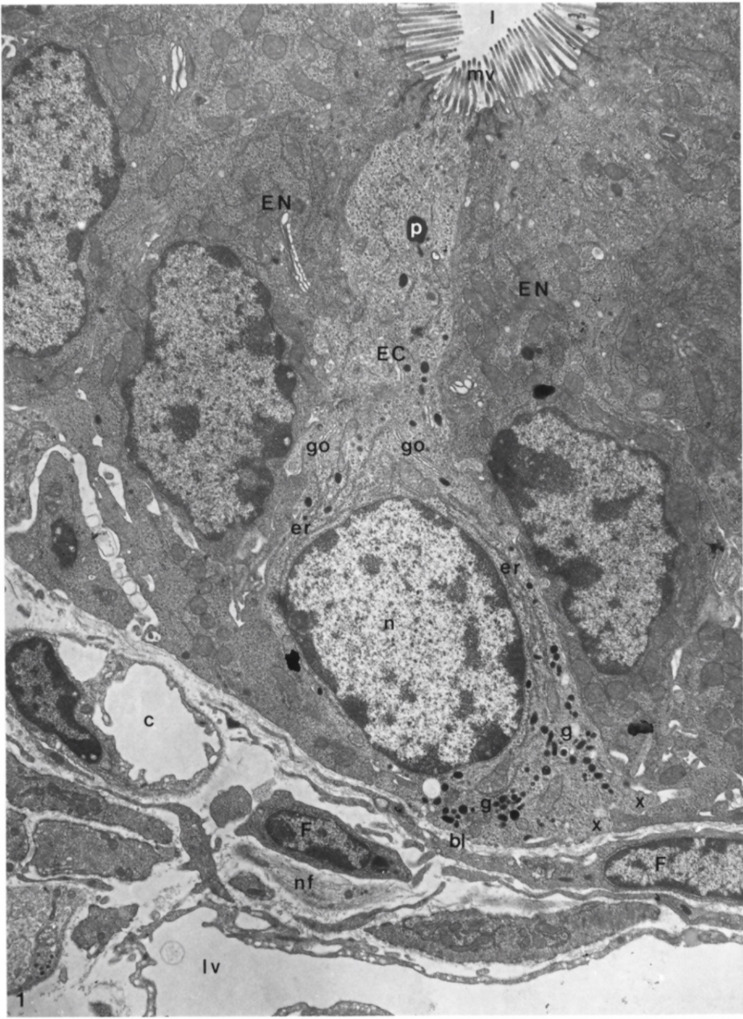
Figure 8.

Longitudinal section of an enterochromaffin cell from the basal lamina to the lumen of a duodenal crypt, in which it is possible to recognize a fenestrated capillary, fibroblast, lymph vessel, and nerve fibers. Moreover, it is also possible to highlight the presence of rough endoplasmic reticulum, secretory granules, Golgi complexes, microvilli, phagosome, and basal cytoplasmic extensions of the enterochromaffin cell. EC: enterochromaffin cell; bl: basal lamina; l: lumen; c: capillary; nf: nerve fibers; lv: lymph vessel; F: fibroblasts; EN: enterocytes granules; g: secretory granules; go: Golgi complexes; mv: microvilli; p: phagosome; x: basal cytoplasmic extension. X 9.600. The Figure is obtained from Wade et al., (1985). Reprinted by permission from Springer Nature Customer Service Centre GmbH: Springer Nature, Cell and Tissue Research (License Number 5232430326152, 19 January 2022) [33].
