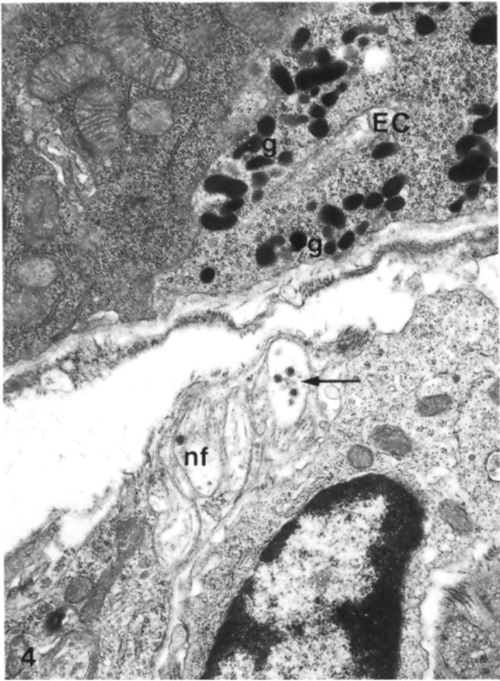
Figure 9.

Base of an enterochromaffin cell fixed in glutaraldehyde and osmium tetroxide in which it is possible to see the presence of secretory granules containing clear and dense-cored vesicles, close to nonmyelinated nerve fibers. EC: enterochromaffin cell; nf: nonmyelinated nerve fibers; g: secretory granules; black arrows indicate dense-cored vesicles. X25.500. The Figure is obtained from Wade and Westfall, (1985). Reprinted by permission from Springer Nature Customer Service Centre GmbH: Springer Nature, Cell and Tissue Research (License Number 5232430326152, 19 January 2022) [33].
