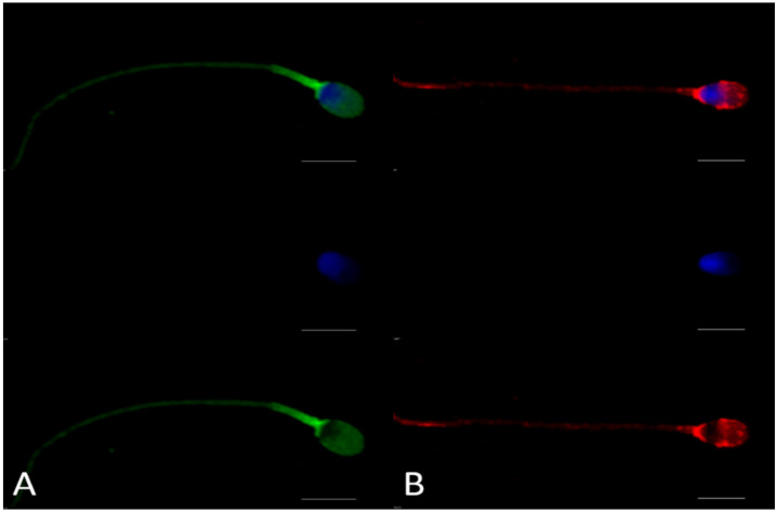
Figure 3.
Immunocytochemistry analysis of ACE2 on ejaculated human sperm cells. Panel (A) shows the location of the C-terminal domain of ACE2 recognized by anti-ACE2-1 (Abcam, ab15348), shared between both isoforms; Panel (B) shows the fluorescence pattern after immunocytochemistry analysis with anti-ACE2-2 (Novus, NBP2-67692), recognizing the full-length ACE2 only. DAPI was used to stain the nuclei. Top: two-laser image (anti-ACE2 antibody + DAPI); middle: sperm cells stained only with DAPI; bottom: sperm cells illustrating only ACE2 fluorescence patterns (scale bar = 5 µm). Negative controls for immunocytochemistry assays are shown in Supplementary Figure S1.

