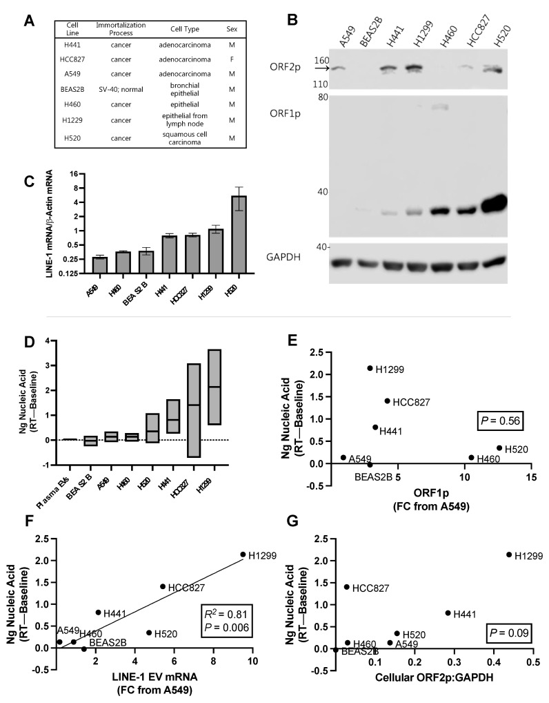
Figure 7.
Relationship between Long Interspersed Element-1 (LINE-1) components and reverse transcriptase activity (RT) in extracellular vesicles (EVs) from lung cancer cell lines. (A) Characteristics of one normal (BEAS2B) and six cancer cell lines used in the experiment. (B) Cellular ORF1p and ORF2p expression. Representative Western blot using 75 μg protein depicting quantities of cellular ORF1p and ORF2p. The ORF2p band is the top band, indicated by the black arrow. (C) Quantification of cellular LINE-1 mRNA levels using One-Step RT-qPCR (Mean, SEM shown). (D) EVs from three independent cultures and EV collections were measured for RT activity (min, max, and mean shown). RT activity from the plasma donors in 6F are shown for reference (“Plasma EVs”). We then quantified LINE-1 components in EVs using an ORF1p ELISA and One-Step RT-qPCR. These measures were normalized to A549s, which exhibited the lowest levels of ORF1p and LINE-1 mRNA. As LINE-1 RT activity requires the formation of a ribonucleoprotein comprised of its proteins and mRNA, we compared RT activity to the levels of each of these components to determine if one of them could be used as a predictor of RT activity (R2 and p levels shown in each figure (E,F). As ORF2p in EV isolates was not detected by Western blotting, we compared EV RT activity with mean cellular ORF2p levels, as approximated by the densitometric analysis of ORF2p Western blots (G). N = 3.

