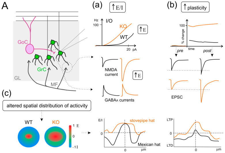
Figure 5.
The IB2 KO mouse model as an example of increased E/I balance, hyper-plasticity, and altered spatial organization of activity in ASD. (A) Schematic view of the granular layer (GL) microcircuit, with mossy fibers (MF) inputs, granule cells (GrC), and Golgi cells (GoC). Three main panels describe the alterations observed in the IB2 KO mouse model of ASD. (a) Increased excitatory/inhibitory (E/I) balance: the additional panels show the input-output (I/O) relationship in granule cells, the NMDA component of excitatory postsynaptic currents in response to MF stimulation, and inhibitory postsynaptic currents, in both WT (black) and KO (orange) conditions. (b) Enhanced long-term potentiation (LTP): the additional panels show the time-course of excitatory postsynaptic currents (EPSC) percent change before and after LTP induction, and EPSC traces pre- and post-induction, for both WT and KO conditions, as in (a). (c) Altered spatial distribution of activity in the granular layer: the additional panels show the “classic” organization in center/surround structures, with excitation prevailing in the core and inhibition in the surrounds. This organization shifts from the Mexican hat to the stovepipe hat profile. Interestingly, this alteration is preserved after plastic changes in synaptic activity, where LTP and long-term depression (LTD) organize mirroring the E/I profile. ((a–c) panels are drawn from the results shown in [370]).

