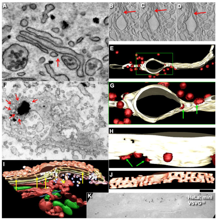
Figure 12.
Pores separate cisternal distensions, which are filled with VLDLs (A) in hepatocytes and procollagen I in human fibroblasts (B–J). (A–D) All distensions are separated from the rest of the cisternae by pores (red arrows) around cisternal distensions. (E,G,H) Three-dimensional model of the Golgi cisterna during the mini-wave. Green arrows, COPI-coated buds (red) on the rim of the pore near the distension. (F) Mini-wave protocol. Tangential section of the medial Golgi cisterna. Immuno EM, enhanced nanogold particles show PC. Red arrows, pores surrounding PC aggregate (black blob) in the section. Asterisks indicate solid parts of Golgi cisternae. (I,J) Three-dimensional model of the Golgi complex with the PC-containing cisterna distension in the perforated cis-most cisterna at 2 min after the transport block. Yellow arrows, many pores in the medial cisternae. (K) HeLa cells. Mini-wave of VSVG. Low level of penetration of VSVG into the Golgi stack. Scale bars (nm): 165 (A); 420 (B–D); 240 (E,F); 75 nm (H); 280 (I,K).

