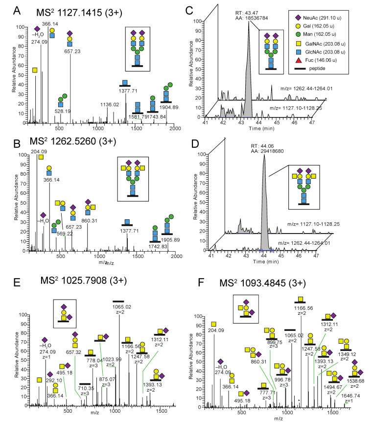
Figure 4.
Glycoproteomic analysis of N- and O-glycopeptides carrying the Sda epitope or its precursor structure in transfected HEK293 cells. MS2 spectra obtained at NCE 20% for (A) a complex type disialo biantennary N-glycopeptide with the amino acid sequence IVDVNLTSEGK including the Asn-174 glycosite (underlined), from transmembrane 9 superfamily member 3 (TM9S3) and for (B) a glycopeptide with the same amino acid sequence carrying one Sda epitope on each of the two antennae. The measured monoisotopic masses for precursor ions are provided in the panel headings (C,D) Extracted ion chromatograms (XICs) of the two precursor ions demonstrate that (C) the mock-transfected sample contains the complex biantennary glycopeptide but not the Sda epitope glycopeptide, and (D) vice versa is true for the wt B4GALNT2-transfected sample. (E) MS2 spectrum obtained at NCE 20% of a glycopeptide, with the amino acid sequence LAGTESPVREEPGEDFPAAR from transferrin receptor protein 1 (TFR1), carrying the disialo core 1 O-glycan, and (F) a glycopeptide with the same sequence carrying the Sda epitope. The glycosite is at Thr-104 or Ser-106. It should be observed that the measured monoisotopic masses for precursor ions at four decimals are provided in the headings of the MS2 spectra. Displayed m/z values of fragment ions are from the largest isotope peaks, not always from the monoisotopic ion. Thus, delta masses in the figures occasionally differ by ±1 u from calculated values.

