Figure 7.
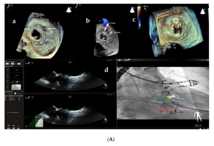
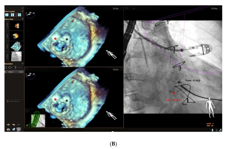
(A) Baseline 3D TEE color Doppler showing two round-shaped mitral PVLs with significant regurgitant jets located at 5 o’clock (3.2 mm) and 7 o’clock (3.6 mm) (a,b) in a female patient with a mitral bioprosthetic valve replacement; (c): successful implantation of a 3 mm-square twist PLD (red asterisk) at 5 o’clock; (d): fusion of real-time 2D TEE (Phillips Epiq7) and cardiac fluoroscopy imaging was obtained using EchoNavigator®-system (Philips Healthcare, Best, The Netherlands) with the fused image maintained demonstrating the location of the postero-laterally located (7 o’clock) mitral PVL (yellow circle) that was marked to aid the pathway of the guidewire (black arrow) crossing the leak, particularly in the case of a radiolucent bioprosthetic valve like in our patient. “*” refers to 3 mm-square twist PLD. (B) Intraprocedural navigation using fusion of 3D TEE and fluoroscopy with the fused image maintained helped the interventionalist to close successfully the second leak with a 5 mm-square twist PLD (black asterisk), improving the safety and the efficacy of this technically challenging procedure. PLD, Occlutech Paravalvular Leak Device ST, square twist, “*” refers to 5 mm-square twist PLD.


