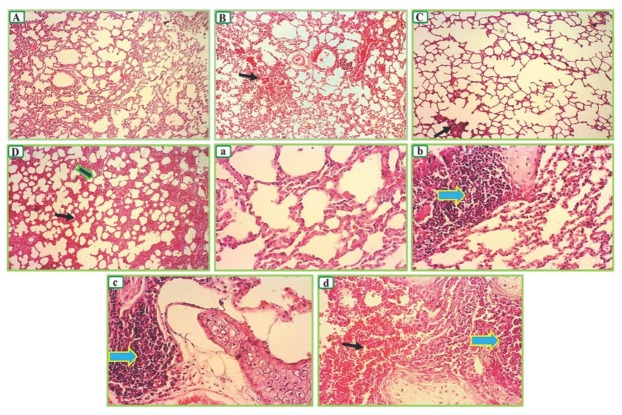
Figure 7.
Representative appearances of the lung tissue stained with H&E. The photographs were taken at 10× (A, B, C, and D) and 40× (a, b, c, and d) magnification. The control group (A and a) had a normal structure. Nevertheless, mild alveolar dilation appeared in noise, toluene, and simultaneous exposure groups. Moreover, imperceptible emphysema appeared in the toluene exposure group. Symbols denote parenchymal hemorrhage (narrow black arrow), inter-alveolar septal thickening (thick black arrow with green outline), and lymphocyte infiltration (thick blue arrow with yellow outline)

