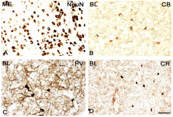
Figure 6.
Brightfield photomicrographs illustrating the immunoreactivity of cellular structures of neuron-specific nuclear protein (NeuN, (A)), calbindin (CB, (B)), parvalbumin (PV, (C)) and calretinin (CR, (D)) of wild-type in the mice amygdala. (A): The medial (ME) and (B–D): The basolateral (BL). Scale bar = 50 µm.

