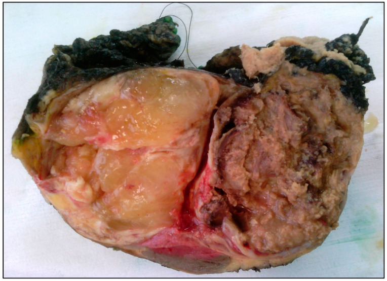
Figure 2.
Macroscopic image of a pediatric low-grade myxoid liposarcoma, excised after neoadjuvant radiotherapy. On the left side of the specimen is a smooth, gelatinous area (characteristically seen in tumors of a lower grade). The right side of the specimen shows necrosis, as a result of the preoperative radiotherapy.

