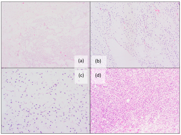
Figure 3.
Histopathological features of myxoid liposarcoma: (a) Characteristic alveolar and edema-like growth pattern (H&E staining); (b,c) Myxoid stroma with arborizing “chicken-wire” vessels and lipoblasts (H&E staining); (d) High-grade MLPS with more than 5% cellular overlap, diminished myxoid matrix, less apparent capillary vasculature, a higher nuclear grade and increased mitotic activity (H&E staining).

