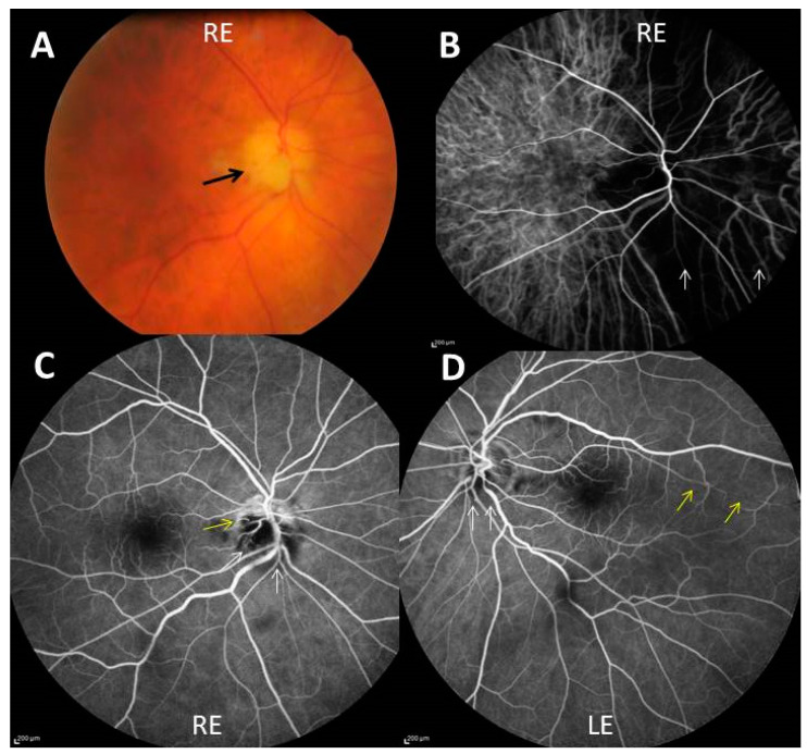
Figure 1.
(A) Chalky white optic disc edema (black arrow) of the right eye (RE); (B) indocyanin green angiography showing severe choroidal hypoperfusion of a large nasal part of the right eye (white arrows); (C,D) fluorescein angiography showing non perfusion of two-thirds of the optic disc (white arrows), outside an upper crescent, and cilio-retinal artery occlusion (yellow arrow) in the right eye (C) and blurring of the lower optic disk margin (white arrows) and hypoperfusion of a long cilioretinal artery (yellow arrows) in the left eye (LE) (D).

