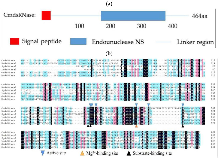
Figure 2.
Analyses of the amino acid sequence of CmdsRNase (dsRNase from C. medinalis). (a) Schematic diagram of the domain of CmdsRNase. The red box, blue box, and blue line represent the signal peptide, endonuclease_NS domain, and linker region, respectively. (b) Multiple sequence alignment of dsRNases from different insect species: CmdsRNase (C. medinalis), BmdsRNase (B. mori, BAF33251.1), SgdsRNase (S. gregaria, AHN55088.1), LmdsRNase (L. migratoria, ARW74135.1), OndsRNase (Ostrinia nubilalis, MT524712.1), and McdsRNase (Mamestra configurata, HM357845.1). Blue inverted triangles represent active sites of six key amino acid residues, the yellow triangle indicates Mg2+-binding sites, and black triangles mark substrate-binding sites.

