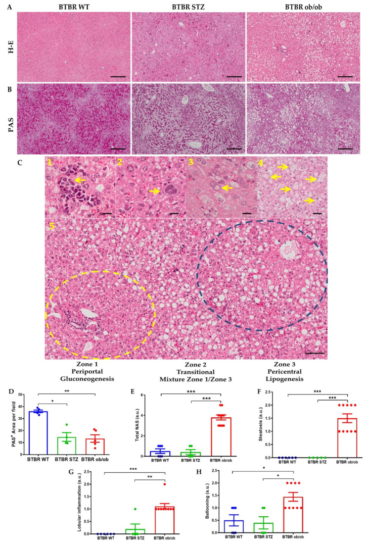
Figure 4.
Liver histopathological changes at 22 weeks between BTBR WT, BTBR-STZ and BTBR ob/ob. Representative images of H-E (A) and PAS (B) staining in the 3 animal groups. (C) Major findings associated to MAFLD development: 1. Inflammatory clusters; 2. Isolated megakaryoblast; 3. Glycogenated nuclei; 4. Hepatocellular ballooning. 5. Steatosis distribution into 3 distinctive liver zones. (D) Quantification of positive Periodic Acid Schiff (PAS) staining, as glycogen liver deposition in BTBR WT, STZ and ob/ob mice. (E) Quantification of NAFLD activity score (total NAS) and its histopathological characteristics: (F) steatosis, (G) lobular inflammation and (H) hepatocytes ballooning. Data are shown as scatter dot plots and mean ± SEM of each group (n = 5–8 mice/group); * p < 0.05, ** p < 0.01, *** p < 0.001 vs. BTBR WT or BTBR-STZ.

