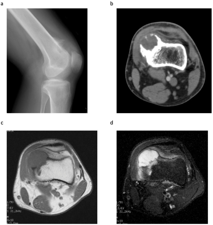
Figure 1.
A 23-year-old male with a pathological diagnosis of periosteal chondrosarcoma (Case 3). (a) Lateral radiograph of the left knee showing a lobulated calcified mass on the anterior aspect of the distal metaphysis of the femur. (b) CT showing the medial periosteal-based lesion with a calcified shell. The cortex is thickened, but there is no evidence of medullary invasion. (c) T1-weighted MRI showing a lobulated mass (6.0 × 4.0 × 3.0 cm), with low signal intensity, arising from the periosteum. (d) T2-weighted fat suppressed MRI showing a predominantly high signal intensity mass with medullary invasion. Adjacent soft tissue oedema is also present.

